| CROSS-DISCIPLINARY PHYSICS AND RELATED AREAS OF SCIENCE AND TECHNOLOGY |
 |
|
|
|
|
Quasi-Two-Dimensional Diffusion in Adherent Cells Revealed by Three-Dimensional Single Quantum Dot Tracking |
| Chao Jiang1,2, Bo Li1, Shuo-Xing Dou1,2, Peng-Ye Wang1,2,3*, and Hui Li1,4* |
1Beijing National Laboratory for Condensed Matter Physics and Laboratory of Soft Matter Physics, Institute of Physics, Chinese Academy of Sciences, Beijing 100190, China
2School of Physical Sciences, University of Chinese Academy of Sciences, Beijing 100049, China
3Songshan Lake Materials Laboratory, Dongguan 523808, China
4School of Systems Science, Beijing Normal University, Beijing 100875, China
|
|
| Cite this article: |
|
Chao Jiang, Bo Li, Shuo-Xing Dou et al 2020 Chin. Phys. Lett. 37 078701 |
|
|
|
|
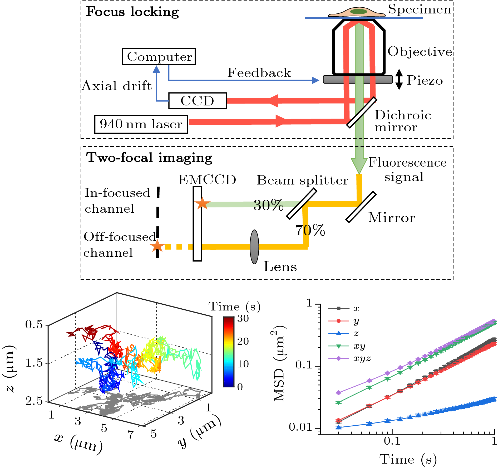
Abstract Intracellular diffusion is critical for molecule translocation in cytoplasm and mediates many important cellular processes. Meanwhile, the diffusion dynamics is affected by the heterogeneous cytoplasm. Previous studies on intracellular diffusion are mainly based on two-dimensional (2D) measurements under the assumption that the three-dimensional (3D) diffusion is isotropic. However, the real behaviors of 3D diffusion of molecules in cytoplasm are still unclear. Here, we have built a 3D single-particle tracking (SPT) microscopy and studied the 3D diffusion of quantum dots (QDs) in adherent A549 cells. Notably, we found that the intracellular diffusion of QDs is quasi-2D, with the axial motion being severely confined. Further investigations demonstrated that disrupting the cytoskeleton component or endoplasmic reticulum (ER) does not alter the quasi-2D diffusion pattern, although ER reduces the diffusion rates and slightly relieves the constraint in the axial diffusion. The preferred quasi-2D diffusion is quite robust and attributed to the complex cytoarchitectures in the flat adherent cells. With the aid of 3D SPT method, the quasi-2D diffusion in cells was revealed, shedding new light on the physical nature of cytoplasm.
|
|
Received: 02 June 2020
Published: 12 June 2020
|
|
|
|
|
| Fund: This work was supported by the National Natural Science Foundation of China (Grant Nos. 11674383, 11874415, 21991133, and 11774407), the National Key Research and Development Program (Grant No. 2016YFA0301500), the Youth Innovation Promotion Association of CAS (Grant No. 2019006), and the Fundamental Research Funds for the Central Universities (Grant No. 2019NTST26). |
|
|
|
| [1] | Luby-Phelps K 1999 Int. Rev. Cytol. 192 189 |
| [2] | Novak I L, Kraikivski P and Slepchenko B M 2009 Biophys. J. 97 758 |
| [3] | Li H et al. 2015 J. Am. Chem. Soc. 137 436 |
| [4] | Lidke D S and Wilson B S 2009 Trends Cell Biol. 19 566 |
| [5] | Luo L and Tang L H 2014 Chin. Phys. B 23 070514 |
| [6] | He W et al. 2016 Nat. Commun. 7 11701 |
| [7] | Verkman A S 2002 Trends Biochem. Sci. 27 27 |
| [8] | Katayama Y et al. 2009 ChemPhysChem 10 2458 |
| [9] | Shen H et al. 2017 Chem. Rev. 117 7331 |
| [10] | Li B et al. 2018 Proc. Natl. Acad. Sci. USA 115 12118 |
| [11] | Pinaud F, Clarke S, Sittner A and Dahan M 2010 Nat. Methods 7 275 |
| [12] | Welsher K and Yang H 2014 Nat. Nanotechnol. 9 198 |
| [13] | Thompson M A et al. 2010 Proc. Natl. Acad. Sci. USA 107 17864 |
| [14] | Risco C et al. 2014 Annu. Rev. Virol. 1 453 |
| [15] | Valm A M et al. 2017 Nature 546 162 |
| [16] | Wells N P et al. 2010 Nano Lett. 10 4732 |
| [17] | Ram S et al. 2008 Biophys. J. 95 6025 |
| [18] | Holtzer L, Meckel T and Schmidt T 2007 Appl. Phys. Lett. 90 053902 |
| [19] | Gosse C and Croquette V 2002 Biophys. J. 82 3314 |
| [20] | Speidel M, Jonas A and Florin E L 2003 Opt. Lett. 28 69 |
| [21] | Wu M M, Roberts J W and Buckley M 2005 Exp. Fluids 38 461 |
| [22] | Sbalzarini I F and Koumoutsakos P 2005 J. Struct. Biol. 151 182 |
| [23] | Toprak E, Balci H, Blehm B H and Selvin P R 2007 Nano Lett. 7 2043 |
| [24] | Diezmann A V, Shechtman Y and Moerner W E 2017 Chem. Rev. 117 7244 |
| [25] | Okada C Y and Rechsteiner M 1982 Cell 29 33 |
| [26] | Li H, Duan Z W, Dou S X and Wang P Y 2012 Acta Phys. Sin. 61 068701 (in Chinese) |
| [27] | Li H et al. 2012 PLOS ONE 7 e45465 |
| [28] | Liu J, Li B H and Chen X S 2017 Chin. Phys. Lett. 34 050201 |
| [29] | Dupont A et al. 2013 New J. Phys. 15 075008 |
| [30] | Etoc F et al. 2018 Nat. Mater. 17 1048 |
| [31] | Kusumi A et al. 2012 Annu. Rev. Cell Dev. Biol. 28 215 |
| [32] | Mogilner A and Manhart A 2018 Annu. Rev. Fluid Mech. 50 347 |
|
|
Viewed |
|
|
|
Full text
|
|
|
|
|
Abstract
|
|
|
|
|
